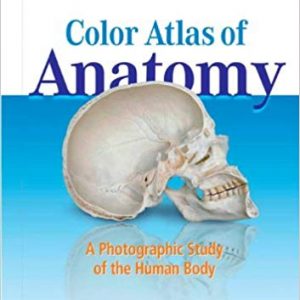
This updated Anatomy: A Photographic Atlas 8th edition (PDF) features outstanding full-color photographs of actual cadaver dissections with accompanying diagnostic images and schematic drawings. Depicting anatomic structures more realistically than illustrations in traditional atlases, this proven resource shows college students exactly what they will see in the dissection lab. Chapters are organized by region in the order of a typical dissection with each chapter presenting topographical anatomical structures in a very systemic manner.
The latest 8th edition features additional clinical imaging such as CTs, MRIs, and endoscopic techniques, as well as new images, graphics, including clinically relevant vessel and nerve varieties and antagonistic muscle functions. Many older images have been replaced with new, higher-resolution images and black-and-white dissection photographs have been replaced with colour photography.
NOTE: This only includes the ebook Anatomy: A Photographic Atlas 8e in PDF. No access codes included.


Reviews
There are no reviews yet.